Vitamin D is one of the oldest hormones on Earth and plays an essential pleiotropic role in maintaining global homeostasis [1]. Although the physiological role of vitamin D is most commonly associated with the maintenance of the musculoskeletal system, the biological properties of this relatively simple compound extend far beyond the regulation of calcium and phosphorus homeostasis [2]. In the past few decades, the extraskeletal effects of vitamin D have become more evident, including its roles in regulating cell proliferation, differentiation, and apoptosis and in immune modulation [3]. The main source of vitamin D is skin exposure to sunlight. Human skin serves both as the site of vitamin D synthesis and as a target organ for its biologically active form [4].
1.1. Vitamin D Synthesis and Functions
The epidermis is one of the most important sources of vitamin D for the body (Figure 1). Upon exposure to sunlight, i.e., ultraviolet (UV) radiation (action spectrum 290–315/280–320 nm or UVB), in the photochemical reaction, 7-dehydrocholesterol (7-DHC) is converted into pre-vitamin D3 in keratinocytes of the basal and spinous cell layers of the epidermis [3,4,5]. Pre-vitamin D3 is then converted into vitamin D3 (cholecalciferol) through thermal isomerization [6]. Following cutaneous synthesis, vitamin D3 enters the circulation primarily bound to vitamin D binding protein (VDBP). In contrast, after intestinal absorption, vitamin D3 is transported in association with both VDBP and lipoproteins [7]. Whether synthesized in the skin or acquired through the diet, vitamin D3 is biologically inactive and undergoes two subsequent hydroxylation reactions to gain its full hormonal activity. First, in the liver, the enzyme vitamin D 25-hydroxylase (CYP2R1) converts it into 25-hydroxyvitamin D (25(OH)D), also known as calcidiol. Then, in the kidney, the enzyme 1α-hydroxylase (CYP27B1) converts it into the active metabolite 1,25-dihydroxyvitamin D (1,25(OH)2D), also referred to as calcitriol. Both 25(OH)D and 1,25(OH)2D can be metabolically deactivated through hydroxylation by the enzyme 24-hydroxylase (CYP24A1) [4]. Vitamin D nutritional status is evaluated by measuring the serum 25(OH)D level, which is the predominant vitamin D metabolite in the bloodstream [5,8].
Aside from producing Vitamin D, keratinocytes, key cells in the epidermis, contain the enzymes CYP27A1 (25-hydroxylase) and CYP27B1 (1-hydroxylase), which are responsible for converting vitamin D3 into its active form, 1,25(OH)2D. Keratinocytes are the only cells in the body that can synthesize vitamin D3 from its precursor, 7-DHC, and convert vitamin D3 to its active metabolite, 1,25(OH)2D [9]. In parallel, several extracutaneous tissues, including the breast, colon, brain, ovaries, and prostate, also express 25-hydroxylase, enabling local conversion of vitamin D3 into 25(OH)D and supporting autocrine production of this metabolite within those tissues [7]. Keratinocytes, like many other cells, express the vitamin D receptor (VDR), allowing them to respond to locally produced 1,25(OH)2D in both an autocrine and paracrine manner [9]. 1,25(OH)2D is a high-affinity ligand of the VDR, which regulates the expression of hundreds of target genes by binding to the vitamin D response elements on the chromosome [10,11]. VDR is most active in keratinocytes during cellular differentiation and proliferation, regulating epidermal proliferation in the basal layer and promoting the gradual differentiation of keratinocytes as they form the upper layers of the epidermis [4,12].
Although initially not considered physiologically significant, the parent compound vitamin D3 plays a crucial role not only in classical endocrine functions but also in the autocrine, paracrine, and intracrine mechanisms of vitamin D actions [7]. While it is well established that vitamin D3 must be metabolized to 1,25(OH)2D to exert its biological effects, the conventional metabolic model has not fully recognized the importance of cellular accessibility to both the parent compound vitamin D3 and its primary circulating metabolite 25(OH)D [7]. Compared to 25(OH)D, vitamin D3 is more effectively internalized by most cell types, except those expressing the megalin–cubilin system, such as cells in the kidney and parathyroid gland. Most other cell types depend on their autocrine or paracrine environments; however, the availability of circulating 25(OH)D is limited due to its strong binding affinity for VDBP. In contrast, the parent compound vitamin D3 has a 10-to-12-fold lower binding affinity for VDBP, allowing a greater proportion of unbound (“free”) vitamin D3 to enter cells and undergo intracellular activation. This difference enhances the bioavailability of vitamin D3 for local conversion to its active form, particularly in tissues with limited access to tightly VDBP-bound 25(OH)D. Enhanced cellular access to vitamin D3 is functionally significant. Following oral administration of vitamin D3, keratinocytes have been shown to upregulate antimicrobial peptides, such as cathelicidin, even after modest rises in circulating 25(OH)D concentrations, suggesting that vitamin D3 itself reaches the skin and is metabolized in situ [7].
1.2. Vitamin D and Epidermal Proliferation and Differentiation
The epidermis consists of four layers of keratinocytes, each at a different stage of differentiation [4]. Epidermal differentiation is a complex, sequential, tightly regulated process (Figure 2) [13]. The basal layer, or stratum basale, rests on the basal lamina (the layer that separates the dermis from the epidermis) and consists of cylindrical keratinocytes along with stem cells whose primary function is the production of transient amplifying cells, which begin the differentiation process as they ascend into the spinous layer [4,12]. Keratinocytes in the basal layer primarily express the keratin pair keratin 5 and 14 [4]. The stratum spinosum, the layer above the stratum basale, contains cells that initiate the production of the keratins K1 and K10, which are characteristic of more differentiated layers of the epidermis. Moreover, precursor molecules of the cornified envelope, such as involucrin, appear in the stratum spinosum, along with the transglutaminase K, an enzyme responsible for the ε-(γ-glutamyl) lysine cross-linking of these substrates into the insoluble cornified envelope [4]. Above the stratum spinosum is the granular layer, or stratum granulosum, which is characterized by electron-dense keratohyalin granules that contain profilaggrin, a precursor of filaggrin, and loricrin, which is an essential component of the cornified envelope. Filaggrin facilitates keratin filament aggregation and, when metabolized into smaller fragments, is also believed to contribute to the hydration of the permeability barrier. Filaggrin mutations are often found in patients with atopic dermatitis [4]. Lamellar bodies, containing both long-chain fatty acids and antimicrobial peptides from the stratum granulosum, line the membrane that separates the stratum granulosum from the stratum corneum [14]. Lamellar bodies are positioned to release their contents into the extracellular space, where the lipids and antimicrobial peptides play a role in maintaining the permeability barrier and defending against microorganisms [15].
As keratinocytes transition from the stratum granulosum to the stratum corneum, they undergo destruction of their organelles with further maturation of the stratum corneum into an insoluble, highly resistant structure that surrounds the keratin–filaggrin complex and is associated with an extracellular lipid milieu [4].
1,25(OH)2D, through its receptor VDR, regulates every step of the differentiation process. In vivo and in vitro studies have demonstrated that vitamin D affects keratinocyte proliferation and differentiation in a dose-dependent manner. Interestingly, low concentrations (≤10−9 m) of vitamin D promote keratinocyte proliferation in vitro, whereas high concentrations (>10−8 m) inhibit proliferation and promote differentiation [16,17]. 1,25(OH)2D promotes the production of keratin 1, involucrin, and transglutaminase in the stratum spinosum, which helps maintain proper barrier function [12]. Furthermore, it induces the synthesis of filaggrin, loricrin, antimicrobial peptides, and long-chain fatty acids and the formation of the cornified envelope in the stratum granulosum [4,12,18]. The process of epidermal differentiation and its regulation by 1,25(OH)2D and VDR is sequential, with different genes and pathways being activated in keratinocytes at various stages of differentiation. VDR and CYP27B1 are expressed throughout the epidermis, with the highest level of expression in the stratum basale [19]. The transcriptional activity of VDR is tightly regulated by a number of co-regulators that can show cell specificity [11,20]. The major co-activator complexes in the epidermis regulating VDR function are the Mediator complex (previously known as DRIP or VDR interacting proteins), of which MED1 is the major VDR-binding component, and the Steroid Receptor Coactivator (SRC) complex, of which SRC2 and SRC3 are the major VDR-binding components [21]. MED1 and SRC3 are expressed in a reciprocal pattern in the epidermis. MED1 is primarily present in the stratum basale, while SRC3 is localized in more differentiated layers, specifically the stratum granulosum [21]. In undifferentiated keratinocytes, MED directly interacts with VDR to inhibit its activity, whereas in the differentiated cell layers of the stratum granulosum, co-activators SRC2 and SRC3 regulate VDR activity, enhancing its function [22]. The knockdown of MED1 in the epithelium leads to increased keratinocyte proliferation and disrupts the expression of keratins 1 and 10 and involucrin [18]. In contrast, SRC3 knockdown reduces glucosylceramide production and impairs lamellar body formation [22]. Both co-activators enhance vitamin D-induced transcription in proliferating cells and are differentially involved in vitamin D-driven differentiation of epidermal keratinocytes. SRC3 regulates terminal differentiation, while MED1 is involved in the regulation of proliferation and early keratinocyte differentiation [21].
In addition to its role in epidermal differentiation, 1,25(OH)2D enhances the innate immune function of keratinocytes by stimulating the expression of toll-like receptor (TLR2) and its co-receptor CD14. This activation triggers a feedback loop in which TLR2 and CD14 induce CYP27B1 expression, leading to the increased production of 1,25(OH)2D. In turn, this promotes the synthesis of cathelicidin, a potent antimicrobial peptide [23,24].
It is important to highlight the role of calcium gradient in the keratinization process. In vivo, there is a gradient of increasing intracellular Ca2+ concentration across the epidermal layers, with the peak concentration occurring in the stratum granulosum [25]. Calcium concentration below 0.07 mM promotes keratinocyte proliferation, whereas acutely raising extracellular calcium levels above 0.1mM (calcium switch) induces the rapid redistribution of several proteins from the cytosol to the membrane, where they participate in epidermal differentiation. These proteins include the calcium receptor (CaR), phospholipase C-γ1(PLC-γ1), Src kinases, various catenins such as β-catenin, phosphatidyl inositol 4-phosphate 5-kinase 1α (PIP5K1α), and the formation of the E-cadherin/catenin complex (adherens junctions) with phosphatidyl inositol 3 kinase (PI3K). These proteins play important roles in calcium and vitamin D-induced epidermal differentiation [26,27,28]. Within hours of the calcium switch, keratinocytes begin producing keratins K1 and K10, followed by increased levels of profilaggrin, involucrin, and loricrin. These proteins, along with others, are cross-linked into insoluble cornified envelope by the calcium-induced transglutaminase, marking the final step in the differentiation process [29].
The CaR is crucial for these calcium-induced responses, and its expression is upregulated by 1,25(OH)2D, which increases the keratinocytes’ sensitivity to calcium prodifferentiation effects [30]. Deletion of CaR in keratinocytes disrupts their response to extracellular calcium by reducing calcium stores and inhibiting the formation of the E-cadherin/catenin complex [26,27]. Mice without the CaR exhibit a defective permeability barrier caused by the abnormal production of essential lipids and proteins necessary for barrier formation, along with an impaired innate immune response. Furthermore, CaR deletion leads to the reduced expression of VDR and CYP27B1, likely contributing to the impaired epidermal differentiation observed in CaR-deficient mice [31]. As mentioned earlier, 1,25(OH)2D induces CaR, and just as 1,25(OH)2D/VDR stimulates CaR expression, calcium/CaR is essential for VDR and CYP27B1 expression. This highlights the strong interaction between calcium and vitamin D signaling in epidermal differentiation. Consequently, 1,25(OH)2D/VDR enhances the keratinocyte response to calcium’s prodifferentiation effects, while CaR’s role in the regulation of VDR and CYP27B1 expression further supports the prodifferentiation actions of 1,25(OH)2D [31]. The synergism between CaR and VDR is clearly demonstrated by their joint regulation of the expression of several genes, including a member of the PLC family [32]. All members of the PLC family are induced both by 1,25(OH)2D and calcium, and inhibition of PLC-γ1 expression blocks the differentiation induced by 1,25(OH)2D and calcium [32]. Another significant synergistic effect of calcium and 1,25(OH)2D in epidermal differentiation is their ability to induce involucrin and transglutaminase, which is due to the close proximity of the calcium response element and vitamin D response element in the involucrin promoter [33].
Understanding the regulatory pathways and molecular mechanisms through which vitamin D influences the skin is essential. Human skin is inseparably connected to vitamin D, serving both as the site of synthesis and a primary target for its action. In this review, we provide a comprehensive overview of current knowledge regarding the role of vitamin D in skin inflammatory diseases, including atopic dermatitis, acne vulgaris, psoriasis vulgaris and hidradenitis suppurativa.
2. Vitamin D and Psoriasis
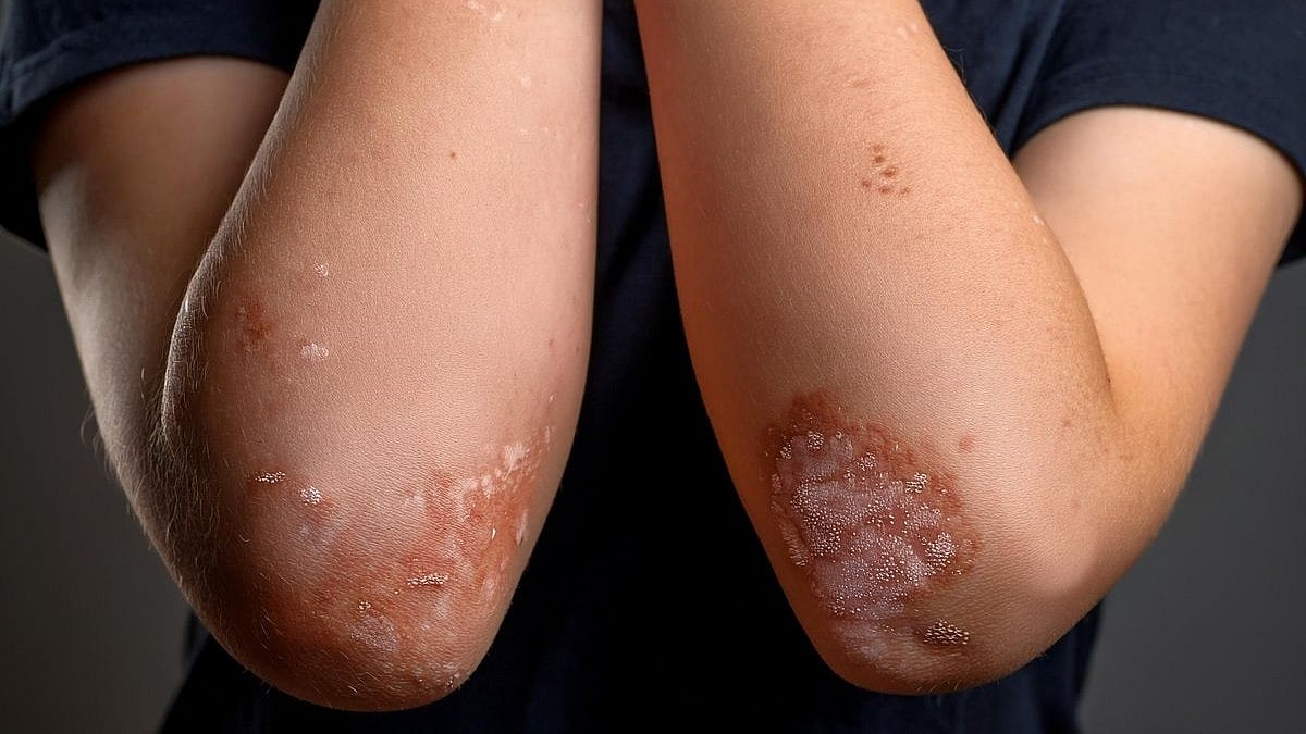
Psoriasis is a chronic, immune-mediated inflammatory disorder affecting about 2–3% of the global population [34]. It is caused by immune dysfunction that leads to continuing inflammation, increased proliferation of skin cells, and the formation of erythemato-squamous plaques [34]. Around 80% to 90% of cases are plaque-type psoriasis, but there are also less common forms like guttate, pustular, inverse, and erythrodermic. Psoriatic patches typically appear on the scalp, elbows, knees, hands, feet, nails, and genitals. They are commonly associated with itching and thickened or pitted nails [35]. An overactive immune system plays a central role in the development of skin lesions [35,36]. Although the exact cause is not completely clear, psoriasis is thought to result from a combination of genetic predisposition and environmental triggers like infections, skin injuries, stress, smoking, or UV exposure [36]. In people with a genetic tendency toward psoriasis, these factors can activate the immune system, leading to inflammation and excessive skin cell growth [37]. On a cellular level, psoriasis is linked to activated T cells and a disrupted balance between Th1 and Th2 immune responses [38]. In healthy individuals, Th1 cells release pro-inflammatory cytokines like IL-2, IFN-γ, and IL-12, while Th2 cells are more involved in anti-inflammatory responses, producing IL-4 and IL-10. In patients with psoriasis, the immune balance shifts toward a Th1-dominant state, leading to an overproduction of cytokines such as IL-2 and IFN-γ, along with a rise in Th17 and Th22 cell activity [36,38]. Th17 cells produce cytokines such as IL-6, IL-17, and IL-22, which help drive inflammation and rapid skin cell growth seen in psoriasis, leading to the development of plaques [36,38]. Moreover, innate immune cells like dendritic cells, macrophages, neutrophils, natural killer cells, and even keratinocytes also play a significant role in psoriasis by producing proinflammatory cytokines, which help intensify the immune response [36]. When dendritic cells are activated by self-DNA and peptides such as LL-37, they produce interferons, which cause the inflammatory response and enhance Th1 and Th17 activity. The inflammation causes keratinocyte hyperproliferation and leads to immune cell migration and neoangiogenesis, forming new blood vessels in affected skin areas [36]. Pro-inflammatory cytokines like TNF-α, IL-17A/F, and IL-22 play a major role in psoriasis. Therefore, treatments that target these molecules have transformed therapy for moderate-to-severe cases. Biologic drugs that block IL-17, IL-23, or TNF-α suppress inflammation by interrupting the signals that promote keratinocyte proliferation and immune system overactivation [36].
The Association Between Vitamin D and Psoriasis
Vitamin D, particularly in its active form 1,25(OH)2D, plays a significant role in immune modulation and skin homeostasis [4]. Vitamin D is known to act on both the innate and adaptive immune systems, so it plays a valuable role in treating psoriasis. Active vitamin D promotes the function of immune cells such as dendritic cells, T-cells, and macrophages by enhancing their phagocytic property and stimulating the production of antimicrobial peptides, such as cathelicidin, which contribute to the body’s defense against infections [39]. Within the adaptive immune system, vitamin D helps reduce inflammation by lowering the activity of Th1 cells by blocking the production of IL-2 and IFN-γ and promoting Th2 cell differentiation. This shift supports the release of anti-inflammatory cytokines like IL-4 and IL-10, which help reduce the immune response [40]. Vitamin D regulates keratinocyte differentiation and proliferation by activating VDR, which supports the production of important barrier proteins like loricrin and filaggrin [36,41]. This supports skin integrity and helps slow the rapid cell turnover that occurs in psoriatic plaques. Hosomi et al. were the first to show that 1,25(OH)2D encourages keratinocyte differentiation by inhibiting DNA synthesis [42]. This led to a reduced number of cells, increased cell size and density, differentiation into squamous, enucleated cells, and enhanced cornified envelope formation.
Topical vitamin D and its analogues are widely used as first-line treatments for mild to moderate psoriasis due to their effectiveness and safety profile [43]. Calcipotriol, introduced in the late 1980s, continues to be one of the most widely used treatments for psoriasis. Interestingly, it has been shown to reduce IL-6 expression in psoriatic lesions, yet it appears to have little or no effect on TNF-α levels, suggesting a more targeted mechanism of action. When used alongside treatments such as UVB phototherapy, fumaric acid esters, cyclosporine, or acitretin, calcipotriol has been found to improve overall therapeutic outcomes [44,45,46,47]. Clinical trials have demonstrated that the fixed-dose combination of calcipotriene (0.005%) and betamethasone dipropionate (0.064%) is highly effective, offering improved skin clearance with few adverse effects [48]. Tacalcitol is another vitamin D analogue that is both effective and well tolerated in long-term treatment. Extended application of ointments containing 4–20 µg/g of tacalcitol has shown significant improvements in PASI scores, as well as good patient tolerance and stable hormonal and calcium levels [49]. Tacalcitol enhances the effect of NB-UVB phototherapy, highlighting its usefulness as a supportive treatment [50]. Maxacalcitol, a more potent vitamin D3 analogue, has demonstrated greater efficacy than calcipotriol and tacalcitol in laboratory settings, promoting keratinocyte differentiation and limiting cell proliferation, avoiding hypercalcemia [51,52]. Trials have verified its therapeutic importance across different dosages, with even better outcomes when paired with NB-UVB [53]. However, the potential for hypercalcemia is higher compared to other analogues, which may limit its use in routine care [54]. The regulatory role of 1α,25-dihydroxyvitamin D3 in cell proliferation and keratinocyte differentiation was observed in three separate in vitro studies [42,55,56].
Several studies have demonstrated a consistent association between low serum 25-hydroxyvitamin D (25(OH)D) levels and psoriasis [57,58,59,60,61,62,63]. For example, Chandrashekar et al. and Maleki et al. found that individuals with psoriasis had notably reduced concentrations of 25-hydroxyvitamin D compared to healthy controls. These findings also revealed a negative association with PASI scores, suggesting that lower vitamin D levels are linked to more severe disease manifestations [57,60]. Bergler-Czop et al. and Pokharel et al. had similar findings in patients with psoriasis [58,61]. These data were further supported by meta-analyses, which consistently show lower 25(OH)D levels in individuals with psoriasis and a strong inverse link to PASI scores [62]. However, not all studies agree; some have found no meaningful differences in vitamin D levels between psoriatic patients and healthy controls [60,63,64]. Upon examining the available research, it remains unclear whether vitamin D deficiency is a contributing factor to the development of psoriasis or a consequence of the disease [65].
The first oral form of vitamin D used in psoriasis treatment was 1α(OH)D, noted for its ability to reduce keratinocyte overgrowth and modify keratin expression [66]. Trials involving higher daily doses (ranging from 5000 to 50,000 IU) have shown encouraging results, with improvements in PASI scores and reductions in inflammatory markers without evident toxicity [67]. Still, the outcomes have not been universally positive. For instance, studies by Ingram et al. and Jarrett et al. using monthly doses of 100,000 IU demonstrated elevated vitamin D levels but failed to produce meaningful clinical improvements [63,64]. Similarly, research by Prystowsky et al. found no additional benefit when oral calcitriol was combined with UVB phototherapy [67]. Gumowski-Sunek et al. observed alterations in calcium metabolism linked to oral calcitriol, a side effect not seen with equivalent topical doses of calcipotriol [68].
The variability in findings across studies may be affected by differences in dose, duration, and formulation of vitamin D analogues. Heterogeneity among followed patients in terms of disease severity, baseline vitamin D status, genetic background, immune function, and presence of comorbidities may also impact the results. For instance, Pokharel et al. identified an inverse relationship between serum vitamin D levels and disease severity, but those correlations did not translate into therapeutic efficacy [61].
Vitamin D is supposed to effect psoriasis through its regulation of keratinocyte proliferation and differentiation, as well as T-cell response. However, in chronic or severe cases, the modulation of T-cell response might not beneficial enough, due to established immune dysregulation and evolved keratinocyte proliferation. Since clinical trials to date are taking in account only individuals with established moderate or severe psoriasis, it leaves a question of potential preventive or early-stage therapeutic effects of vitamin D. This highlights a significant gap in the literature regarding the timing and context in which vitamin D supplementation might offer the greatest benefit.
In summary, although high-dose oral supplementation may offer therapeutic value, topical vitamin D formulations continue to be the safer and more widely favored option. New analogues that specifically target the skin’s vitamin D pathways represent a promising direction for future psoriasis treatments.


Leave a Reply